
Presentation
Acute onset of left knee lateral side pain for the last few days. No trauma/ fever.
Patient Data
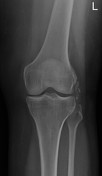
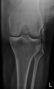
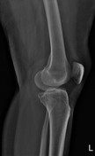
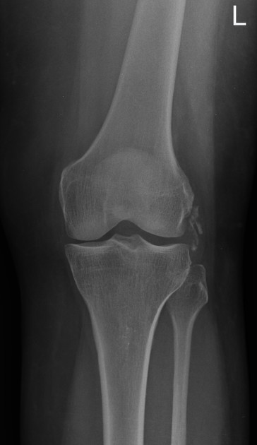
There are a few soft tissue calcification foci in the location of the lateral collateral ligament. There is a mild reduction of the medial femorotibial compartment. There is no fracture/ dislocation/ bone lesion. There is no joint effusion.


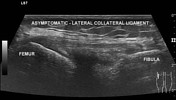
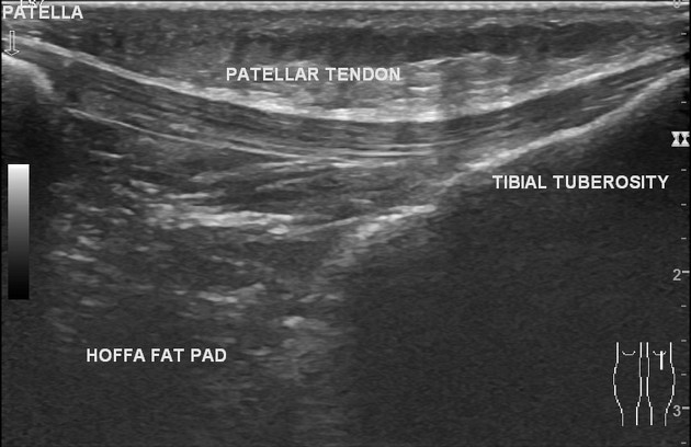
The lateral collateral ligament is intact. There are a few calcification foci (7 - 10 mm) in the cranial half of the ligament. The biceps femoris, popliteus tendon, iliotibial band, quadriceps tendon, and patellar tendon are normal. There is no effusion in the suprapatellar recess. There is no parameniscal cyst.
Case Discussion
The case shows lateral collateral ligament calcification which is an uncommon cause of lateral knee pain.


 Unable to process the form. Check for errors and try again.