
Presentation
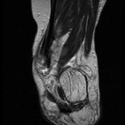
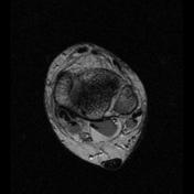
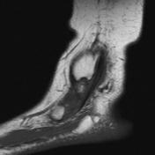
Posterior tibial tendon dysfunction and lateral ankle pain in a patient with valgus deformity of the hindfoot.
Patient Data
Age: 55
Gender: Female


Download
Info
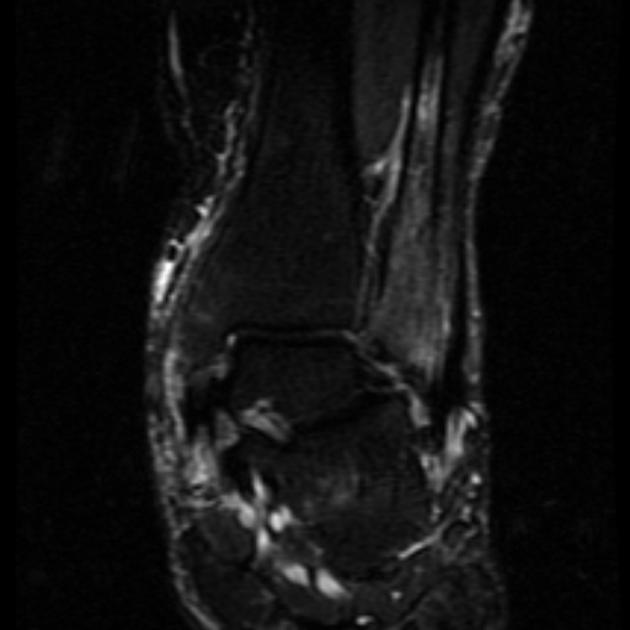
The valgus position of the calcaneus has led to combined talocalcaneal and subfibular lateral hindfoot impingement with edema and cystic changes in the distal fibula and osteoarthritis as well as scar tissue formation between the lateral talus and calcaneus. There are also chronic degenenerative changes of the posterior tibial tendon involving a split tear (best seen on the coronal images) and chronic tenosynovitis with osteophytes along the retrotibial tendon groove. Effusion in the flexor hallucis longus sheath.


 Unable to process the form. Check for errors and try again.