
Presentation
Known rheumatic heart disease with shortness of breath
Patient Data
Age: 20 years
Gender: Female
From the case:
Left atrial enlargement (due to mitral valve regurgitation)

Download
Info

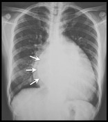
There is cardiomegaly with dilated left atrium and left ventricle.
The upper lung zone vasculature is prominent suggestive of pulmonary venous hypertension.
From the case:
Left atrial enlargement (due to mitral valve regurgitation)

Download
Info

Annotated image demonstrating the double density sign of left atrial enlargement.
From the case:
Left atrial enlargement (due to mitral valve regurgitation)

Download
Info

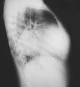
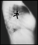
Annotated lateral radiograph shows the markedly dilated left atrium displacing the left main bronchus posteriorly resulting in the "walking man" sign.
Case Discussion
This case demonstrates the radiographic features of chronic mitral valve regurgitation of a patient with rheumatic heart disease. The valve pathology was confirmed on echocardiography.


 Unable to process the form. Check for errors and try again.