
Presentation
Suspicion of a left cervical mass.
Patient Data
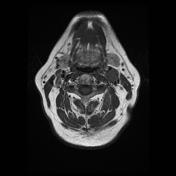

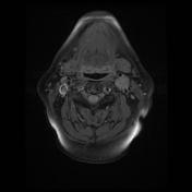
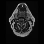
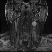
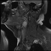

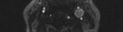
MRI shows a round lesion located between the internal and external carotid bifurcation, iso-intense to muscle on T1 & T2 (salt & pepper appearance), and enhancing avidly after gadolinium administration. The internal and external carotid arteries are splayed by the lesion.
The most probable diagnosis is a carotid body paraganglioma (carotid glomus tumor / chemodectoma)

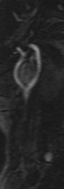
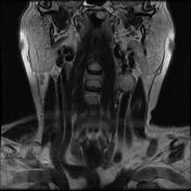
MRI TOF revealed the enhancing left carotid bifurcation lesion, splaying each of carotid artery branches.
Case Discussion
The differential diagnosis is quite limited in this case, as there is a classic salt & pepper appearance of the lesion on T1 weighted images and splaying of the ICA / ECA. This is almost pathognomonic of carotid body paraganglioma.
The differential diagnosis for a tumor in this location includes :
vagal schwannoma: tends to displace both vessels together rather than splaying them
vagal neurofibroma: tends to displace both vessels together rather than splaying them
lymph node mass: may look similar if hypervascular
vagal paraganglioma: same pathology but located more rostrally
carotid bulb ectasia


 Unable to process the form. Check for errors and try again.