
Presentation
Abdominal pain for 1 month, has acutely exacerbated. Elevated lactate.
Patient Data


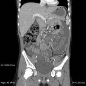
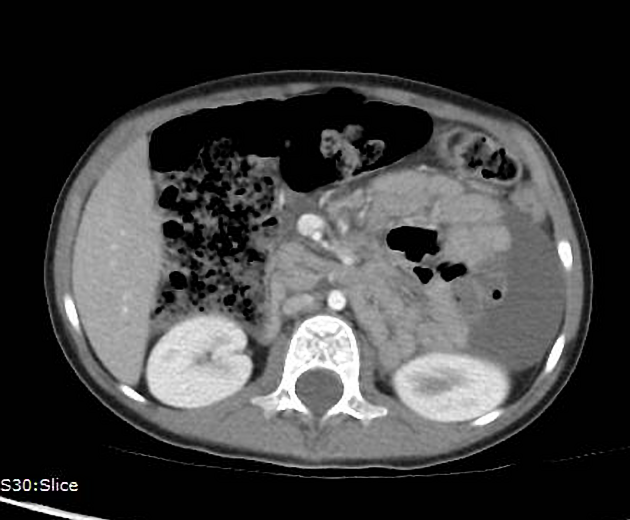
Abnormal location of small bowel loops within the left hemi-abdomen, lying in the left anterior pararenal space. The ectopic small bowel is contained within a hernia sac, which is outlined by ascites. Associated stretching and distortion of mesenteric vessels. Findings are consistent with left paraduodenal hernia.
Additionally, some of the small bowel loops demonstrate severe wall thickening and abnormal enhancement pattern consistent with ischemia in the setting of elevated lactate.
Case Discussion
At surgery, abnormal location of bowel was noted with the additional finding of twisting of the mesentery resulting in venous occlusion and bowel ischemia. After devolvulizing, the bowel was deemed viable and no bowel was resected. Additionally, congenital bands were seen near the ligament of Treitz, which were lysed. Postoperative course was uncomplicated.


 Unable to process the form. Check for errors and try again.