
Presentation
Cough.
Patient Data
Age: 35 years
Gender: Female

Download
Info
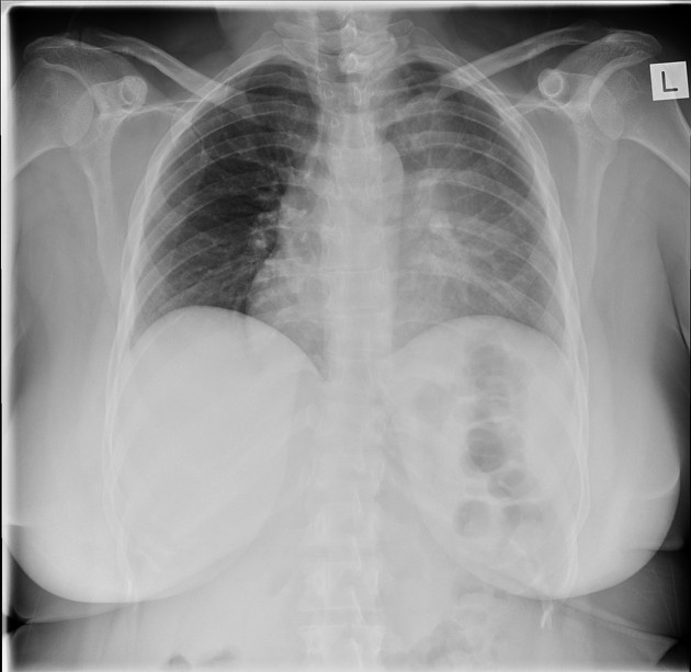
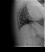
Hazy or veiling opacity extending from the left hilum, obscuring the left cardiomediastinal contour, in keeping with left upper lobe collapse. Luftsichel sign demonstrated.


Download
Info

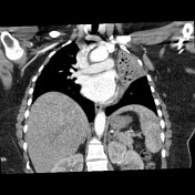
CT confirms left upper lobe collapse, with apparent "cut-off" of the left upper lobe bronchus, which could be due to mucous plugging or other endobronchial lesions.
Case Discussion
Left upper lobe collapse.
Subsequent bronchoscopy shows a pale polypoid endobronchial mass at orifice left upper lobe and discharge of caseous material post-biopsy.
Histology revealed benign stromal elements ? hamartoma. Culture negative to date.


 Unable to process the form. Check for errors and try again.