
Presentation
Presents with left loin pain to groin. Query renal calculus. Past history renal calculus.
Patient Data


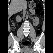
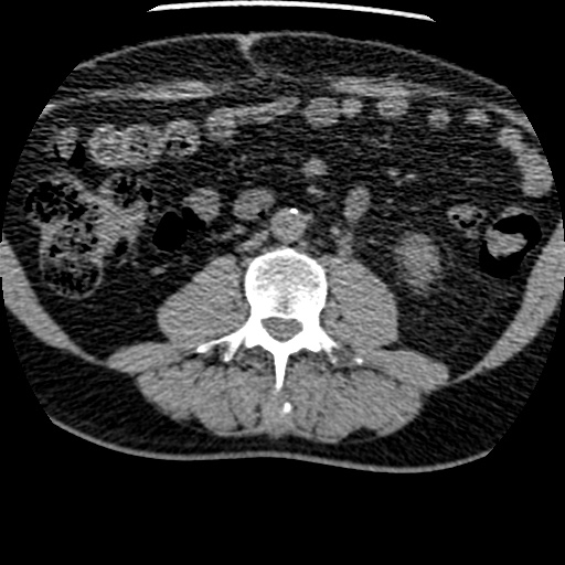
Within the left distal ureter, just proximal to the vesicoureteric junction, there is a 6 x 5 x 5 mm calculus. This is associated with mild left hydroureter and left hydronephrosis, and mild left perinephric stranding. Three small nonobstructing calculi are seen within the left kidney, measuring 3 mm at the upper pole, 2 mm at the lower pole and 4 mm at the lower pole. No right-sided renal calculus identified. Two low density lesions in the liver, measuring 2.6 cm in segment 2/3 and 2.2 cm in segment 6/7, likely represent cysts. Diverticular disease of the sigmoid colon. No dilated loops of small or large bowel.

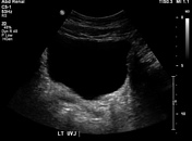
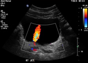
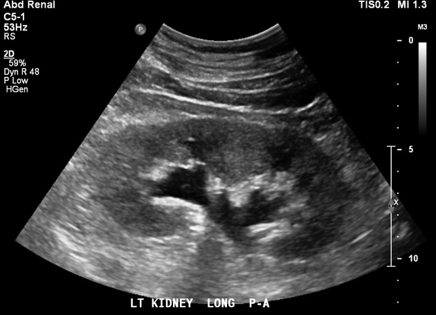
Mild left hydronephrosis. Near the left VUJ, there is a 6mm echogenic focus producing posterior shadowing consistent with a calculus. Only the right ureteric jet was visualized.
Case Discussion
Typical CT and ultrasound appearance of ureteric colic. While non-contrast CT is typically more sensitive for visualizing a ureteric stone than ultrasound, the latter test avoids the need for ionizing radiation and can reliably demonstrate features of obstruction (hydronephrosis, absence of a ureteric jet on color Doppler) even if the stone itself is not seen.


 Unable to process the form. Check for errors and try again.