
Presentation
Patient arrives in the emergency room with pain in the left iliac region with increased white blood cells and C-reactive protein.
Patient Data
Age: 65 years
Gender: Female
From the case:
Lipoleiomyoma


Download
Info
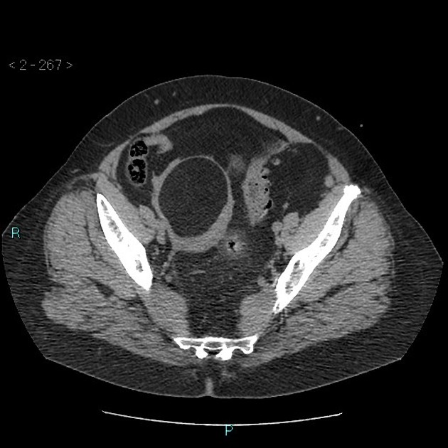
CT without contrast shows sigmoid diverticulitis.
There is also a well-marginated low attenuation intramural lesion in the uterus, compatible with a lipoleiomyoma.
From the case:
Lipoleiomyoma
Download
Info
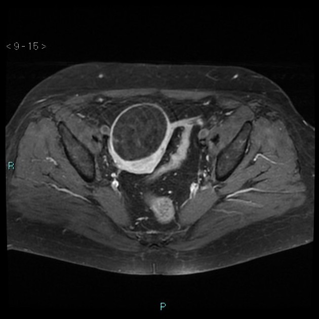
Magnetic resonance imaging with gadolinium shows fat saturation in the region and confirms fat within the uterine lesion.


 Unable to process the form. Check for errors and try again.