
Presentation
Long-standing right anterior chest wall mass. An interval increase is seen in its size in the last couple of years. The patient also complains of mild pain in it which is also a new finding.
Patient Data


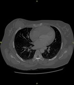

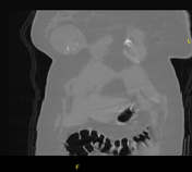
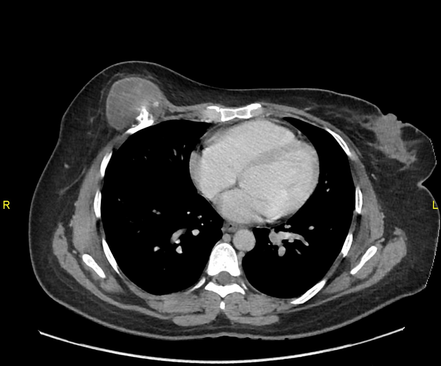
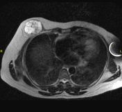
A bony outgrowth/exostosis measuring approximately 2.1 x 1.3 cm is seen along the outer aspect of the anterior right fifth rib, close to the costochondral junction. A well-defined soft tissue component (hypodense to the muscles), measuring approximately 3.7 x 3.7 cm showing no significant enhancement, is seen in relation to this bony exostosis. No underlying rib destruction is noted. Visualized lung fields are clear.

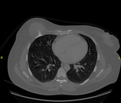
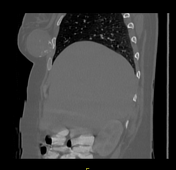
Bony exostosis/osteochondroma is seen along the outer aspect of the anterior end of right 5th rib which is stable when compared with the previous scan; however, an interval increase is seen in the associated overlying soft tissue component (which is likely a cartilaginous cap) which now measures 60 x 45 mm as compared to the previous measurement of 37 x 37 mm. No radiological signs of invasion are seen either into the surrounding soft tissues or underlying ribs. No evidence of local or distant metastasis is seen as well. An interval increase in the size of the cartilaginous cap is suspicious which needs further evaluation with MRI (for the exact thickness of the cap) and possible subsequent histopathological correlation to rule out malignancy.
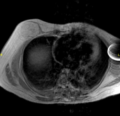
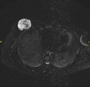
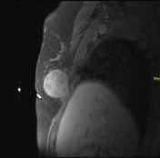
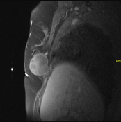
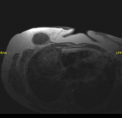

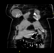
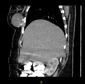
A well-defined mass lesion measuring about 58 x 44 mm is seen along the right anterior 5th rib, just under the right breast. This mass is of high signal intensity on T2 and STIR and isointense to the subcutaneous fat on T1-weighted images. The lesion has a peripheral low signal intensity rim on all pulse sequences, which may represent ossification or calcification. It shows mild enhancement on the post-contrast scan. These MRI features are suggestive of a thick cartilage cap around the osteochondroma and the possibility of malignant transformation into the secondary chondrosarcoma needs to be ruled out.
Case Discussion
The lesion was completely excised and diagnosed as low-grade chondrosarcoma (likely secondary).
Malignant transformation occurs in the cartilage cap of osteochondroma. Cartilage cap thickness of > 1.5 cm or continuous growth of the cap after growth plate closure is suspicious of malignant transformation. Malignant transformation occurs in approximately 1% of solitary osteochondromas, which is generaly of low-grade type and can be managed with surgery in the majority of cases.


 Unable to process the form. Check for errors and try again.