
Presentation
Dyspnea
Patient Data


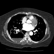
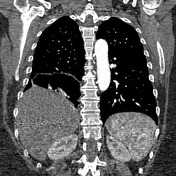
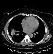
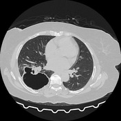
A large intrapulmonary cavity is seen at the posterior aspect of the right lower lobe with air-fluid level and uniform thick wall. It is surrounded by minimal ground-glass densities and atelectasis, suggestive of lung abscess.
Normal course, caliber, enhancing pattern and branching of the main pulmonary trunk, its right and left branches as well as their lobar and segmental branches. No evidence of pulmonary embolism.
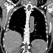
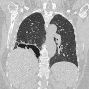

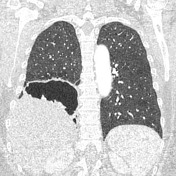
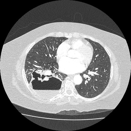
Mild regression in size and wall thickness of the previously noted right lower lobar cavitary lesion. No current evidence of the air-fluid level detected within the cavitary lesion. The surrounding related ground-glass attenuation density and atelectatic plates are still noted.
Case Discussion
Features are suggestive of lung abscess, resolved with antibiotic treatment. Follow up and further investigations are recommended as it has many differential diagnoses like cavitating bronchogenic carcinoma, cavitating pneumonia, and necrotic metastasis.
At presentation, it shows benign features of a pulmonary cavity as uniform wall thickness, absence of mediastinal or hilar lymphadenopathy, and minimal surrounding consolidation with preserved air bronchogram.


 Unable to process the form. Check for errors and try again.