
Presentation
Chest pain, fever and breath shortness.
Patient Data
Age: 20 years
Gender: Female


Download
Info

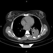
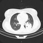
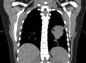
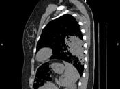
A rounded mass-like lesion, 60mm in diameter, is seen occupying most of the left lower lobe and shows an irregular margin with internal air bubbles. After contrast media injection, mild peripheral enhancement is observed.
In addition, a few enlarged lymph nodes are seen at the left hilar region.
Case Discussion
Pathologically proved lung abscess in a young adult female.
CT is the most sensitive and specific imaging modality to diagnose a lung abscess. Contrast should be administered, as this enables the identification of the abscess margins, which can otherwise blend with surrounding consolidated lung.


 Unable to process the form. Check for errors and try again.