
Presentation
Shortness of breath and fever. Initially treated for pneumonia. Followed 5 days later by sudden right sided pleuritic chest pain and increased shortness of breath.
Patient Data

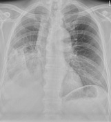
The admission chest radiograph shows dense consolidation affecting the right upper lobe.
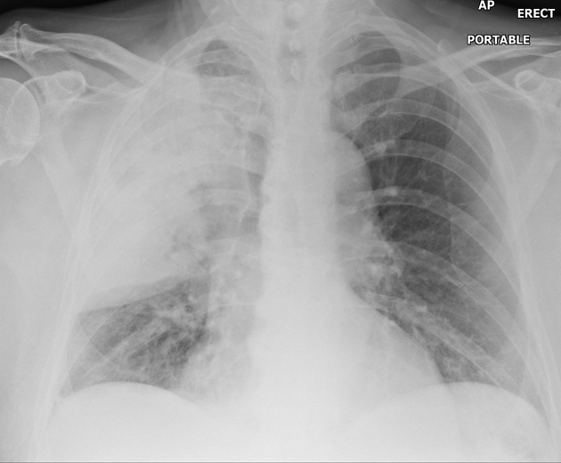
A follow up radiograph at 5 days after presentation showed unexpected new consolidation within the right lower zone with resolution of the changes in the right upper zone.
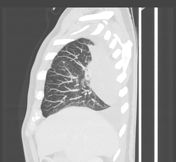
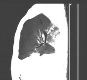

Follow up CT shows a 90 clockwise rotation of the right oblique fissure and a primarily posterior position of the right upper lobe.
Reformatted images reveals twisting of the lobar bronchial anatomy with up-going middle and lower lobe bronchi.
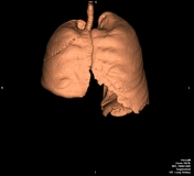
Volume rendering shows the right oblique fissure clearly visible on the anterior surface.
Case Discussion
The patient initially presented with signs and symptoms of a pneumonia with corresponding dense right upper lobe consolidation on chest X-Ray.
Five days later following antibiotic treatment he complained of sudden onset right sided pleuritic chest pain and a worsening of his shortness of breath.
CT revealed the right lung had torsed causing potential threefold compromise of his airways, pulmonary arteries and veins. He was taken to a local cardiothoracic unit where the diagnosis was confirmed and underwent right upper and middle lobectomies.
Lung torsion is a very rare event and is almost invariably associated with a history of previous surgery. In this case it is theorized that an odd rotational force during coughing began a clockwise rotation of the right lung which was amplified by the density of the lobar consolidation.


 Unable to process the form. Check for errors and try again.