
Mechanism of action of adenosine in cardiac MRI perfusion imaging
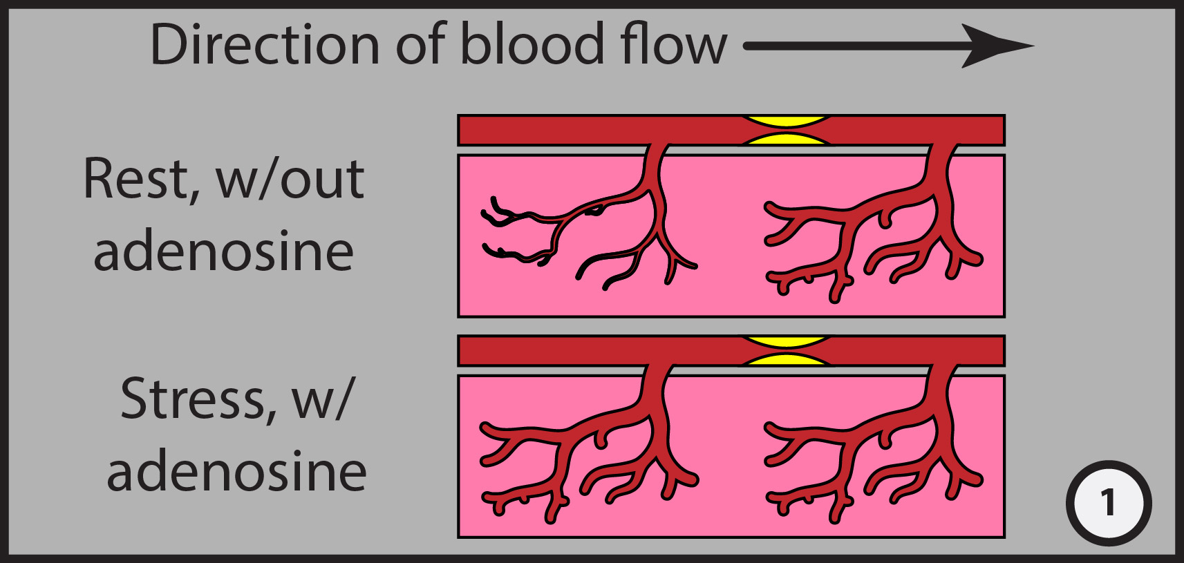
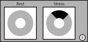
Illustration #1: Myocardial blood vessels are maximally dilated downstream from a severe stenosis, but are normal proximally. With stress, i.e. administration of adenosine, myocardial vessels proximal to an obstruction dilate, but those distal to the stenosis cannot dilate further, resulting in relative decreased perfusion in ischemic myocardium when comparing rest to stress images.
Illustration #2: After IV gadolinium administration, the rest short axis "image" is normal, but the stress image obtained with adenosine shows a perfusion defect. Note the endocardial involvement: defects due to coronary artery disease will always involve the endocardium because the endocardium is most vulnerable to ischemia.
Case Discussion
Original illustrations by Stefan Tigges.


 Unable to process the form. Check for errors and try again.