
Presentation
Headache and confusion. The patient had history of malignant melanoma.
Patient Data
Age: 55 years old
Gender: Male
From the case:
Melanoma metastases to brain


Download
Info
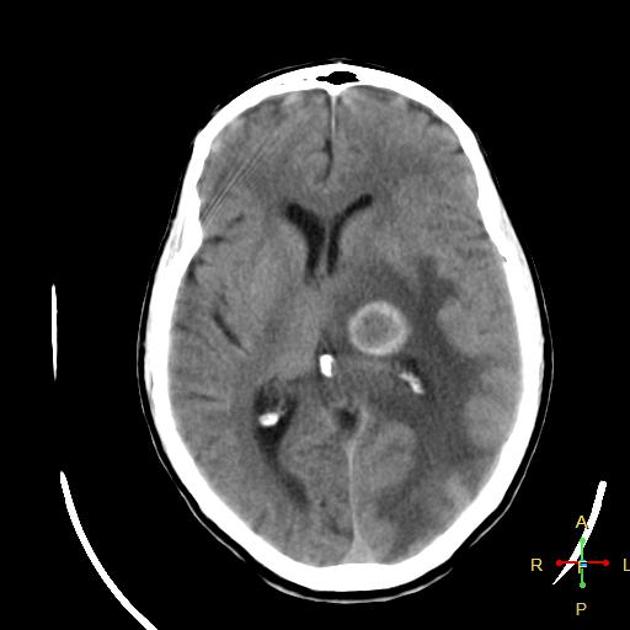
Left occipital, parietal and basal ganglionic space nodules. They are hyperdense in non contrast study and show marginal enhancement in post contrast study, surrounded by perifocal edema that exerts mild contralateral subfalcine herniation.
Case Discussion
Melanoma is the third most common primary neoplasm that metastasizes to the brain.


 Unable to process the form. Check for errors and try again.