
Patient Data
Age: Adult
From the case:
Meningioma - burnt-out

Download
Info
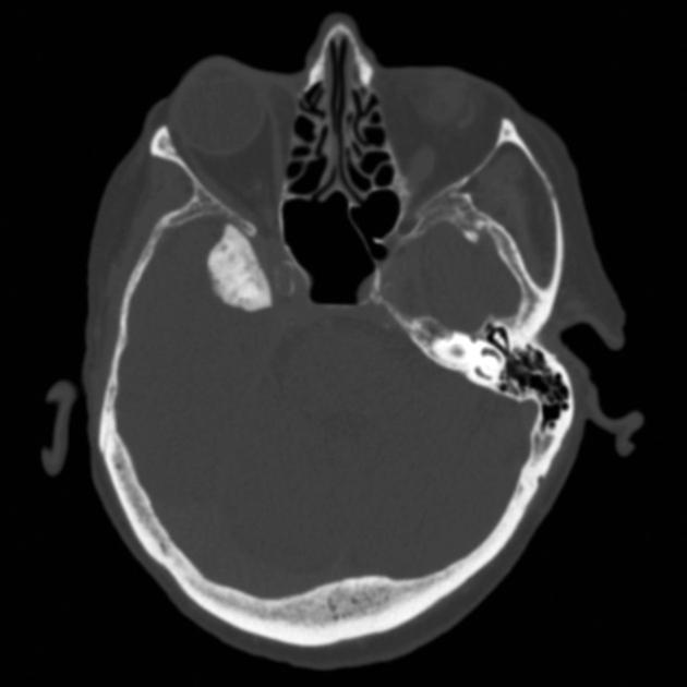
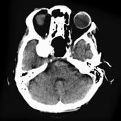
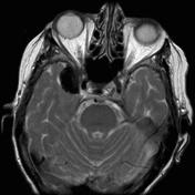
Heavily calcified extra-axial mass in the right middle cranial fossa is consistent of a heavily calcified "burnt out" meningioma.
From the case:
Meningioma - burnt-out
Download
Info
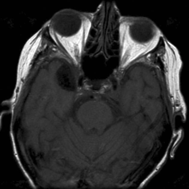
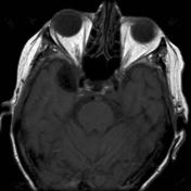
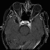
Extra-axial mass is of low signal on all sequences with only slight surrounding enhancement.
Case Discussion
Features are typical and characteristic of a heavily calcified burnt out meningioma.


 Unable to process the form. Check for errors and try again.