
Presentation
Right fourth nerve palsy.
Patient Data


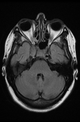
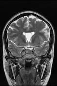
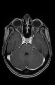
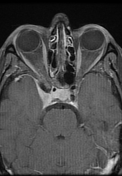

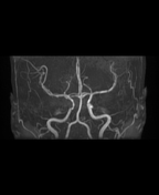
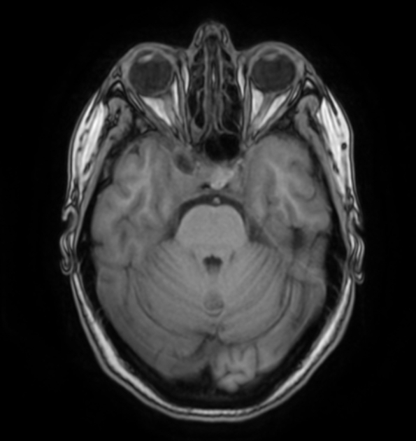
The MRI sequences demonstrate a right parasellar mass (27 x 20 x 17 mm) centered on the cavernous sinus, displaying an isosignal to the cortical grey matter on T1/T2, and FLAIR with vivid homogeneous enhancement on postcontrast sequences and hyperostosis of the anterior clinoid. There is encasement with the reduced caliber of the ipsilateral internal carotid artery. Medially there is an extension to the sellar region with a shifted pituitary stalk to the left. Posteriorly a partial filling of the Meckel cave with mild thickening and enhancement of the prepontine dura are noted.
Cavum septum pellucidum is incidentally noted.
Case Discussion
MRI features are most consistent with meningioma of the cavernous sinus.
On imaging, there is a wide differential (see cavernous sinus mass).


 Unable to process the form. Check for errors and try again.