
Presentation
Imaging follow-up after one year.
Patient Data
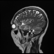
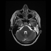

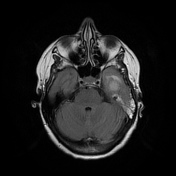
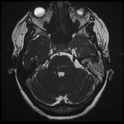
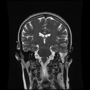
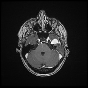
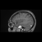
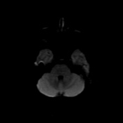
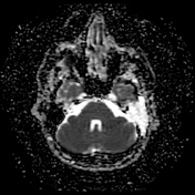

MRI sequences demonstrate a solid extra-axial nodule arising from the floor of the left middle cranial fossa/anterior portion of the petrous bone, showing to be isointense on T1 and slightly hyperintense on T2 compared to the adjacent cortex, with a vivid and homogeneous enhancement and mild diffusion restriction compared to the adjacent cortex. There is a mild mass effect due to the mass projecting into the left temporal lobe from the floor, characterized by local vasogenic edema. The lesion measurements and aspect are overall stable compared to the previous MRI.
Note is made to a diffuse obliteration of the mastoid air cells on the left.
Case Discussion
This case demonstrates an extra-axial tumor arising on the floor of the left middle cranial fossa/anterior portion of the petrous bone, which shows to be stable compared to the previous follow-up imaging. Despite the local mass effect with edema in the left inferior temporal gyrus, the patient was referred to be asymptomatic.
The radiological features are strongly suggestive to a meningioma, and in the context of this patient, no other differential is considered.
Observation is recommended in older patients when the tumor is small and asymptomatic.


 Unable to process the form. Check for errors and try again.