
Presentation
Left orbital pain with reduced visual acuity.
Patient Data


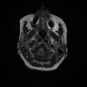
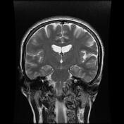
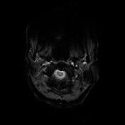
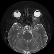
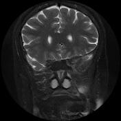
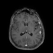

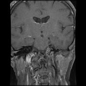
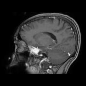
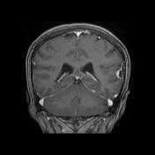
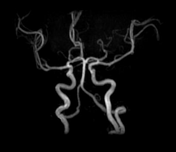

Well-circumscribed extra-axial mass centered on the left cavernous sinus encasing the intracavernous, clinoid and supraclinoid segments of the internal carotid artery. It elicits an isointense signal to cortical grey matter on T1, T2 and FLAIR with no calcification or hemorrhagic component on GE sequence. The postcontrast sequences show a vivid homogeneous enhancement as well as dural thickening with enhancement of the left temporal fossa. There is an intraorbital extension compressing and displacing the optic nerve medially. The MRA 3D-TOF shows reduced caliber from C4 to C7 segment of the left ICA.
Another small broad-based extra-axial enhancing mass is noted in the left temporal lobe, containing foci of calcification with a dural tail sign and adjacent reactive hyperostosis.
Case Discussion
MRI features most consistent with cavernous sinus meningioma encasing the ICA with intraorbital extension compressing and displacing the optic nerve with associated small temporal meningioma.


 Unable to process the form. Check for errors and try again.