
Presentation
Untreatable epilepsy
Patient Data
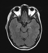

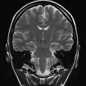

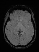
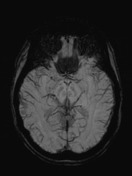

MRI T2 and double inversion recovery (DIR) sequences demonstrate a clear hippocampi asymmetry where the left hippocampus is smaller than the right (hippocampal atrophy) and has an increased T2 signal.
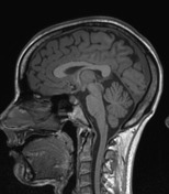
Remaining parenchyma volume is age-appropriate. No other abnormalities identified in the current MRI protocol.
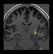
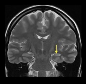
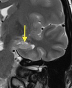

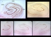
Annotated images demonstrating a left reduced hippocampal volume (arrow) reflecting a hippocampal atrophy, as well an increased T2 signal.
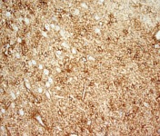
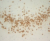

MACROSCOPIC DESCRIPTION: 1. "Hippocampus": A segment of hippocampus with attached mesial temporal tissue - 25x11x7mm. Serially sliced and all submitted. 2. "Normal lateral neocortex": A wedge shaped portion of cerebral cortex and white matter - 30x20x10mm. Serially sliced and all submitted.
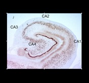
MICROSCOPIC DESCRIPTION: 1. Serial sections of hippocampus show almost complete loss of neurons from the CA1 sector of the hippocampal cortex. There is mild neuronal depletion in the CA3 and CA4 sectors. Neuronal numbers in CA2 are intact. The neuronal loss is accompanied by moderate reactive astrocytic gliosis. There is moderate loss of neurons from the dentate fasciculus with dispersion of residual neurons. The features are of hippocampal sclerosis - ILAE Type 2. There is no inflammation. No evidence of tumlur is seen. 2. The sections show temporal neocortex and white matter. There is moderate gliosis of the sub-pial molecular layer (Chaslin's sclerosis). Cortical lamination is intact. No features of malformation of cortical development are seen. Moderate numbers of neurons are noted in white matter. Myelination is normal. There is no inflammationm and no evidence of tumor is seen.
DIAGNOSIS:
1. Hippocampus: Hippocampal sclerosis - ILAE Type 2.
2. Lateral neocortex: Moderate Chaslin's sclerosis only.
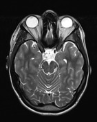
Note: The first image with annotated hippocampal segments corresponds to a normal hippocampus from another patient for comparison.
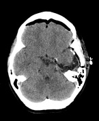
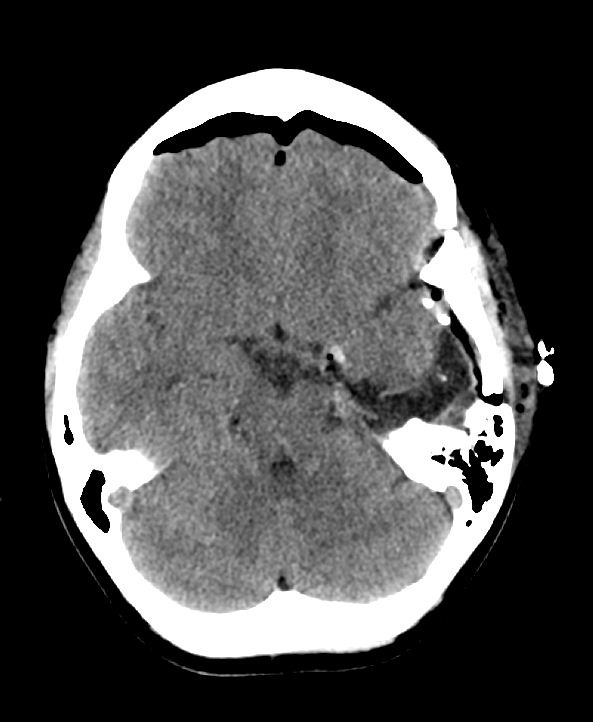
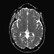
CT scan shows a left temporal craniotomy and left temporal lobectomy. 9 mm hyperdensity along the left tentorium cerebelli is noted, in keeping with acute blood. It is unclear whether this represents extra or intra-axial blood. Further small foci of hyperdensities are seen along the surgical bed. Moderate bilateral pneumocephalus is in keeping with post-operative status. No midline shift. No hydrocephalus. Grey-white matter differentiation is preserved.
Case Discussion
Mesial temporal sclerosis (MTS) is the most common association with intractable temporal lobe epilepsy (TLE).
This case illustrates a typical clinical presentation followed by characteristic MRI features for hippocampal atrophy and further treatment and pathology confirmation.


 Unable to process the form. Check for errors and try again.