Presentation
Chiropractic manipulation for neck pain resulted in altered sensation distal to T11 and saddle anesthesia
Patient Data


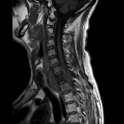
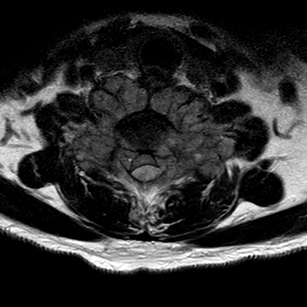
Spinal cord compression and myelopathy.
Large mass infiltrating C7 vertebra with extradural and extraspinal soft-tissue masses. The posterior component of the extradural mass contains higher signal intensity on T1 weighting compatible with hemorrhage. Subtle increased T2-weighted signal intensity in the spinal cord.
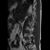
Left upper quadrant mass in the abdomen seen on lumbar spine MR.

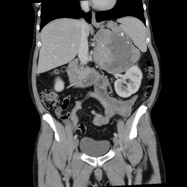
14 x 13 x 11cms adrenal mass with a well-defined lobulated contour, areas of necrosis and calcification, indenting the upper pole of the left kidney and displacing it. No venous invasion or abdominal metastases. Small fluid attenuation liver lesions are likely to be cysts.
Case Discussion
Adrenocortical carcinoma is rare and usually presents late, with mass effect, metastases or Cushing's Syndrome. The diagnosis was made on core biopsy of the cervical mass and confirmed following C7 laminectomy and C5-T2 fusion.


 Unable to process the form. Check for errors and try again.