
Presentation
Worsening left shoulder pain, swelling and weakness over a period of 2 weeks.
Patient Data
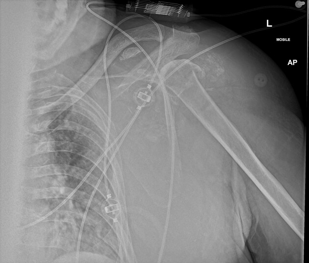
Bony fragmentation of the left humeral head and neck, of indeterminate age, with significant adjacent soft tissue calcification.
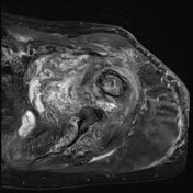

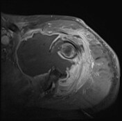
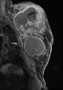

Large shoulder joint effusion with extensive capsular nodular thickening and hyperenhancement with communicating extension into the subacromial recess. The internal contents are heterogeneous with regions of T1 hyperintense signal favored to represent hemorrhage and T2 hypointense regions which may represent debris. Disruption of the all four rotator cuff muscles at the humeral insertion.
The imaging features are not typical for a tumor process with non-enhancing larger heterogeneous fluid collection which is centered on the shoulder joint in association with a proximal glenoid fracture.
Case Discussion
This patient went for ultrasound guided aspiration of the joint effusion which came back positive for hydroxyapatite crystals.


 Unable to process the form. Check for errors and try again.