
Presentation
Pain and swelling left lateral hip.
Patient Data
Note: This case has been tagged as "legacy" as it no longer meets image preparation and/or other case publication guidelines.

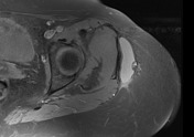
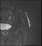
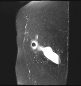

Circumscribed thin-walled fluid collection at the interface between the subcutaneous fat and deep fascia, with fluid-fluid level and small globular focus of fat signal intensity.
Case Discussion
Post-traumatic, closed, internal degloving injuries.
One-third of patients present months or years after the initial injury and can mimic a soft tissue tumor, but the location and shape of the lesion, conforming to that of the fascial plane is atypical for tumor.
Site: usually subcutaneous tissues over the greater trochanter. Also described along flank, buttock, lumbar spine, scapula, and knee.
Macroscopic pathology: oval, fusiform, or crescentic, consistent with fluid dissecting along traumatized fascial planes.
MRI findings: hemolymphatic fluid, blood degradation products, fat (viable or necrotic), capsule variably present, varying degrees of enhancement.
The presence of a capsule may be a factor in choosing surgical treatment over conservative management.


 Unable to process the form. Check for errors and try again.