
Presentation
Antenatal fetal assessment.
Patient Data


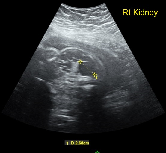
The right kidney is enlarged, reaching right iliac fossa, measuring 6 .4 x 4 cm in dimensions with echogenic dysplastic parenchyma and multiple discrete renal cysts, largest averages 2.7 cm in diameter at the upper pole. No pelvicalyceal dilatation.
Normal configuration of the left kidney, measuring 4.1 x 1.8 cm in dimensions with normally collapsed renal pelvis and calyces, no backpressure changes. No cystic changes. Normal parenchymal echopattern.
Normal filling and sonographic features of the urinary bladder.
Adequate amount of amniotic fluid (single pocket = 6 cm).
Case Discussion
Features are consistent with multicystic dysplastic right kidney, recommended for postnatal ultrasound assessment or fetal MRI. Differential diagnosis includes fetal hydronephrosis, cystic abdominal mass, tuberous sclerosis, and obstructive cystic renal dysplasia. The full bladder and adequate amniotic fluid imply functioning other kidney and no threat on fetal life.


 Unable to process the form. Check for errors and try again.