
Presentation
Acute-onset of left orbital pain with mild proptosis and diplopia.
Patient Data
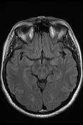

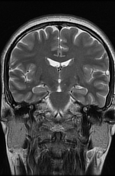
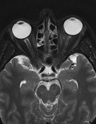
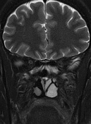
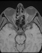

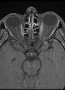
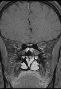
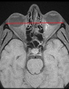

Fusiform enlargement of the left inferior rectus muscle involving its tendinous insertion with axial proptosis grade I (well-visualized on annotated image), and mild inflammatory changes of the adjacent orbital fat. It shows an isosignal to the normal extraocular muscles on T1, high signal on T2 with relatively homogeneous enhancement on postcontrast sequences. Normal appearance of the other extraocular muscles.
Normal appearance of the right orbit.
Mild peripheral mucosal thickening of the maxillary antrum with partial filling of the ethmoid cells.
Case Discussion
MRI features suggestive of a myositic orbital pseudotumor involving the left inferior rectus muscle.
One of the main differential diagnoses of idiopathic orbital inflammation is orbital lymphoma which is usually bilateral with progressive clinical presentation and shows on MRI lower values on ADC and does not respond to corticosteroid.


 Unable to process the form. Check for errors and try again.