
Presentation
Previous lump resection in the thigh.
Patient Data
Age: 25 years
Gender: Male

Download
Info
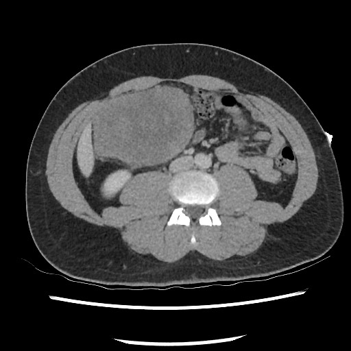
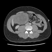
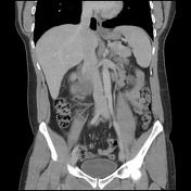
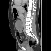
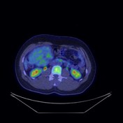
Large and relatively well-defined mass likely in the peritoneal cavity, at the root of the mesentery to the left, showing heterogenous enhancement due to internal areas of low attenuation. No associated lymphadenopathy. The solid and hollow abdominal viscera are normal.
Download
Info
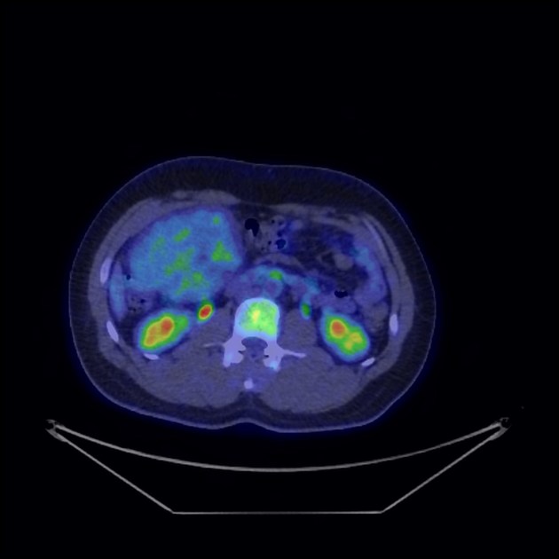
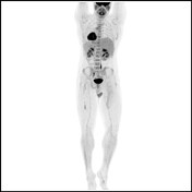
The mass has low, if any, FDG activity.
Case Discussion
This patient has had a myxoid liposarcoma resected a few years ago. Although no prior PET scan available for comparison, these tumors are known to demonstrate low-grade FDG activity.


 Unable to process the form. Check for errors and try again.