
Presentation
Midline cystic swelling.
Patient Data
Age: Child
From the case:
Nasal dermoid


Download
Info
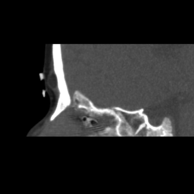
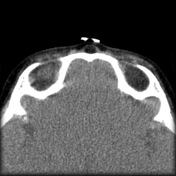
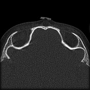
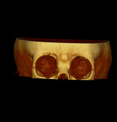
A midline cystic lesion is seen above the level of the nose with central fat density, smooth wall and minimal underlying calvarial scalloping.
From the case:
Nasal dermoid
Download
Info
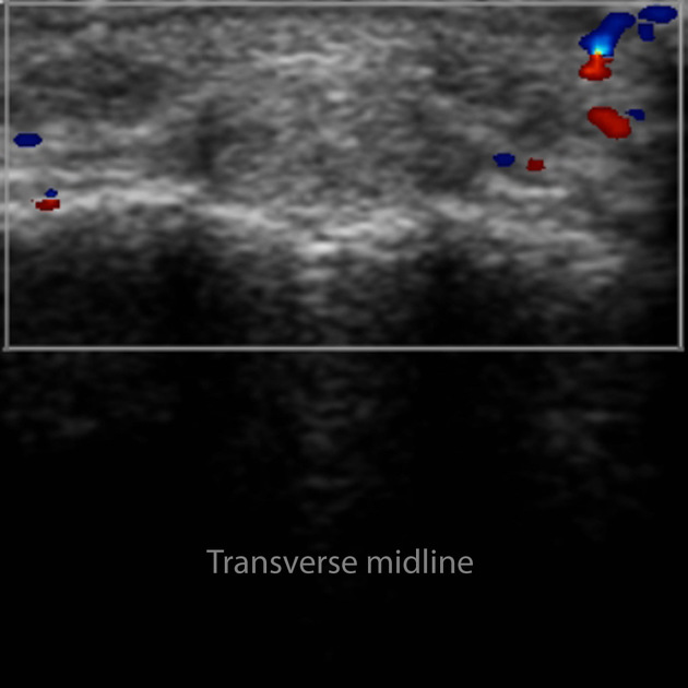
Ultrasound of the lesion shows homogeneous hyperechoic content with no internal vascularity with color Doppler.
Case Discussion
Small midline nasal dermoid in a young child - note the fat density on CT distinguishing it from a nasal epidermoid.


 Unable to process the form. Check for errors and try again.