
Presentation
Respiratory distress
Patient Data
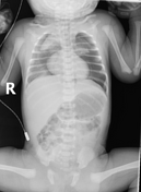

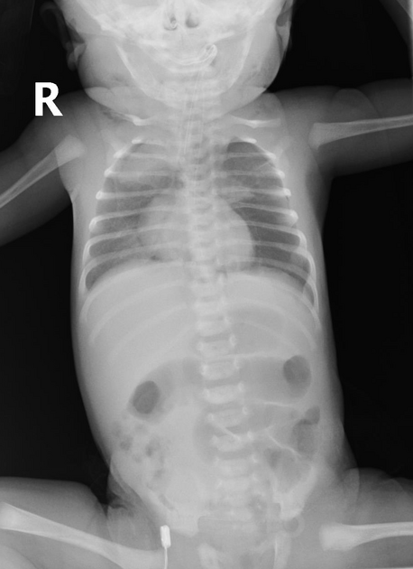
A large amount of air is noted in the anterior and middle mediastinum displaying a spinnaker sign ( Thymus is prominent and outlined by air) suggesting pneumomediastinum.
Moderate subcutaneous emphysema is seen at the base of the neck.
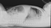
Follow-up after 24 hours shows some improvement in the pneumomediastinum and left pneumothorax .
Case Discussion
Pneumomediastinum is an uncommon cause of neonatal respiratory distress. Although clinical history and examination of the neonate may be uninformative in determining the etiology of respiratory distress, a chest X-ray can be diagnostic of pneumomediastinum.
Chest X-ray shows both thymic lobes lifted and displaced laterally due to the air in the mediastinum. This creates a wedge-shaped opacity extending into the superior mediastinum (spinnaker sing or Angel Wing’ sign).


 Unable to process the form. Check for errors and try again.