
From the case:
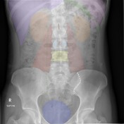
Normal abdominal radiograph - annotated x-ray

Download
Info
Purple: Liver
Pink: Spleen
Green: Left 11th rib
Orange: Kidneys
Red: Psoas muscle
Brown: Spinous process of L1
Light Blue: Pedicles of L3
Black: Transverse processes of L3
Yellow: Vertebral body of L4
Dark Blue: Urinary bladder
Dotted Green: Usual path of the ureter (not usually visible)
Dotted White: Left sacroiliac joint
Case Discussion
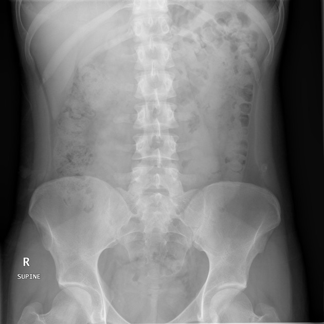
The source image is taken from this case of normal abdominal radiographs.


 Unable to process the form. Check for errors and try again.