
Presentation
Reduced head circumference on second trimester ultrasound.
Patient Data


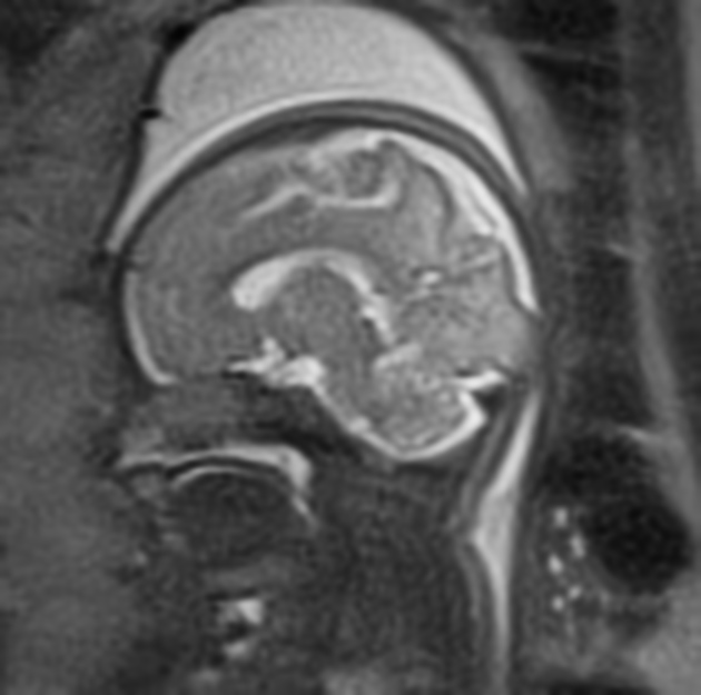
The fetal brain and cerebral biometry were normal. This study demonstrates normal sulcal development at 30 weeks gestation. The operculum is now closed. Superior temporal sulcus and occipitotemporal sulci are now seen and secondary occipital and frontal sulci are developing. Note that the multilayered laminar pattern seen at earlier gestations is no longer present.


Normal fetal body imaging at 30 weeks gestation. Note marked T1 hyperintensity of meconium in colon and thyroid, and slight T1 hyperintensity in liver compared with bowel. The rectum ends well below bladder neck. The adrenal glands are difficult to identify separate from the kidneys. Lung signal intensity on T2 weighted images is approximately twice that of liver.


 Unable to process the form. Check for errors and try again.