
From the case:
Normal hepatic vein spectral Doppler
Download
Info
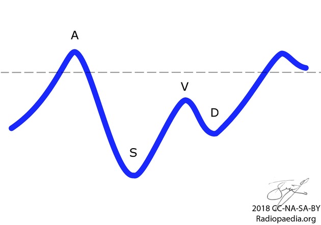
Diagram showing normal hepatic spectral Doppler waveform.
- A wave - atrial contraction causes retrograde flow back to liver, then rapid RV filling
- S wave - ventricular systole, antegrade flow into right atrium against closed TV
- V wave - opening of tricuspid, followed by rush of antegrade flow
- D wave - ventricular diastole, antegrade flow into RV
* RV = right ventricle, TV = tricuspid valve
Case Discussion
For use in article figures.


 Unable to process the form. Check for errors and try again.