
Presentation
First episode of psychosis.
Patient Data
Age: 20 years
Gender: Male

Download
Info

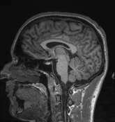
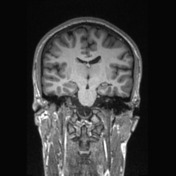
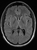
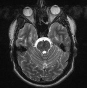
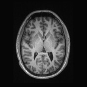
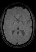
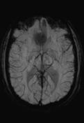
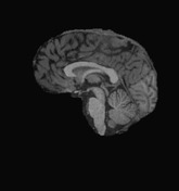
There are no abnormal focal areas of altered signal intensity in the cerebral hemispheres, brainstem or cerebellum. Appearance and intensity of brain parenchyma is normal. Ventricular system and cisternal spaces appear normal. No evidence of intracranial space occupying lesion or obvious vascular anomaly is detected. There is no shift of the midline structures.
Conclusion:
Normal brain study
Case Discussion
This case illustrates a normal brain MRI scan in a neurodegenerative protocol: with a volumetric isometric T1, axial T2 limited to basal ganglia and posterior fossa, axial FLAIR, SWI, and DWI/ADC.


 Unable to process the form. Check for errors and try again.