
Presentation
Pleuritic chest pain.
Patient Data
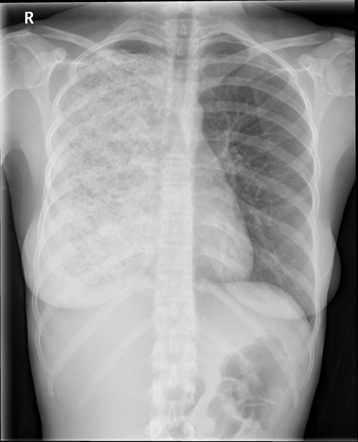
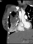
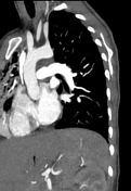
There is extensive air space opacification due to infection involving all segments of the right lung. The appearances of the right hemithorax maybe due to prior surgery for oesophageal atresia and pull-up of the stomach into the right hemithorax.
The left hemithorax is clear.
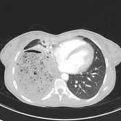

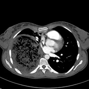
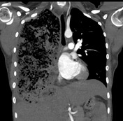

No pulmonary embolism. The mediastinum appears normal. No pericardial effusion. No aortic dissection involving the arch or descending aorta is evident. There is no mediastinal, hilar or axillary lymphadenopathy.
A large collection within the right hemithorax represents stomach (the patient had a gastric pull through procedure as a neonate) and not massive empyema. The stomach does appear rather distended. There are some gastric vessels demonstrated coming up from the coeliac axis. The distended stomach may be related to a recent large meal. However, a gastric outlet obstruction may need to be considered. Clinical correlation suggested, and further investigation indicated as required. There does appear to be a small amount of right-sided pleural fluid adjacent to the stomach. The right lung appears anteriorly compressed. There is consolidation affecting the right lung anteriorly likely related to atelectasis. Pneumonia cannot be completely excluded. There is a shift of the mediastinum to the left.
No endobronchial lesion is seen.
Case Discussion
This patient was diagnosed with oesophageal atresia as a baby. She had successful corrective surgery at that time.
Oesophageal atresia refers to an absence of the continuity of the oesophagus due to an inappropriate division of the primitive foregut into the trachea and oesophagus. it is a condition usually diagnosed in the neonatal period. Some relevant facts about oesophageal atresia include:
- it is the most common congenital anomaly of the oesophagus (~1:3000-4500 live births)
- may be sporadic, but most are found associated with other abnormalities:
- other intestinal atresias: duodenal atresia, jejunoileal atresia, anal atresia
- annular pancreas
- pyloric stenosis
- VACTERL
- CHARGE syndrome
- chromosomal anomalies such as: trisomy 21 and trisomy 18
- it is frequently associated with a tracheo-oesophageal fistula , with the majority of them (~85%) characterised by a proximal atresia with distal fistula


 Unable to process the form. Check for errors and try again.