Presentation
Generalized tonic-clonic seizure.
Patient Data


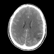
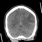
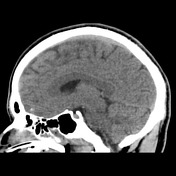

An ill-defined left parietal mass without calcification of convincing enhancing component is present.
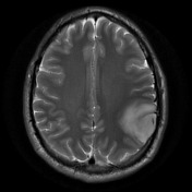
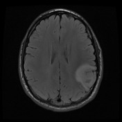
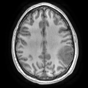
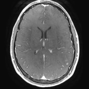
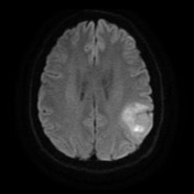
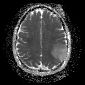
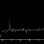
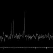

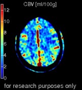
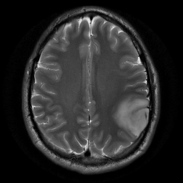
A cortical/subcortical mass demonstrating T2 hyperintense expansion and only minor contrast enhancement is centered in the left inferior parietal lobule. High signal in the DWI sequence is predominantly T2 shine through, although there are small regions of intermediate to low ADC values at the superomedial and anterosuperior aspects. Moderate cerebral blood volume (CBV) increase. Spectroscopy demonstrates choline elevation and NAA reduction.
Conclusion: left inferior parietal lobule mass typical of a low-grade glioma (astrocytoma or oligodendroglioma)
Case Discussion
The patient went on to have surgery.
Histology
MICROSCOPIC DESCRIPTION:
Sections show a moderately cellular tumor composed of moderately pleomorphic cells containing rounded, hyperchromatic nuclei with inconspicuous nucleoli and focal perinuclear clearing. Tumor cells are arranged in diffuse sheets. Focal microcystic change and secondary structuring are observed. No mitotic figures are identified. No necrosis or microvascular proliferation is seen.
IMMUNOHISTOCHEMISTRY:
- GFAP: Positive
- Nogo A: Positive
- Nestin: Positive (low)
- IDH-1 R132H: Positive (mutated)
- ATRX: Positive (non-mutated)
- MGMT: Negative (likely methylated)
- p53: Equivocal
- p16: Positive
Topoisomerase labeling index: Approximately 5%.
FISH for chromosome 1p/19q deletion:
- Chromosome 1.
- Mean copies 1p per cell: 1.12
- Mean copies 1q per cell: 1.6
- 1p/1q ratio: 0.70
- Interpretation: 1p loss detected by FISH
- Chromosome 19.
- Mean copies 19q per cell: 1.13
- Mean copies 19p per cell: 1.73
- 19q/19p ratio: 0.65
- Interpretation: 19q loss detected by FISH
SUMMARY: Chromosome 1p/19q co-deletion detected by FISH
FINAL DIAGNOSIS:
Oligodendroglioma (IDH mutated, chromosome 1p/19q co-deleted) - WHO Grade II.


 Unable to process the form. Check for errors and try again.