
This case has been tagged as "legacy" as it no longer meets image preparation and/or other case publication guidelines.
From the case:
Omental infarction

Download
Info
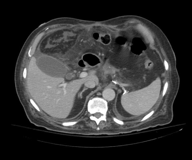
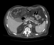
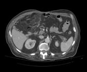
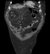
Right upper quadrant large omental heterogeneous lesion of mixed fat and soft tissue densities with adjacent fat plane smudging.
Case Discussion
CT images demonstrate large right upper quadrant omental infarction. Patient was post-operative for esophagectomy with gastric pull-up.
This case was donated to Radiopaedia.org by Radswiki.net


 Unable to process the form. Check for errors and try again.