
Presentation
The patient presented with left sided exophthalmos.
Patient Data
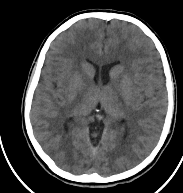
CT show intra-conal focal lesion of fluid density encasing the left optic nerve which appears prominent and tortuous.
Two partially calcified mass lesions in the subcutaneous tissues of the scalp suggesting trichilemmal cysts.
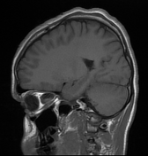
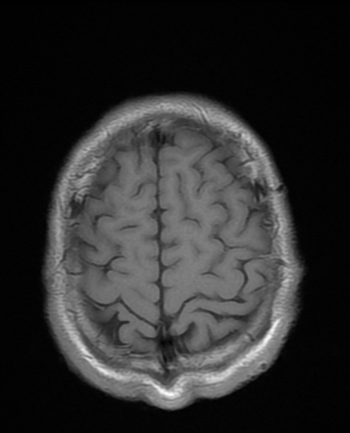
CT brain otherwise normal.


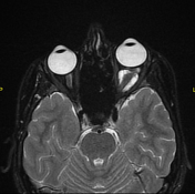

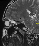
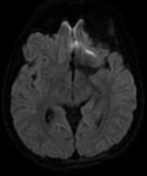
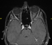
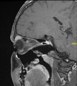
intra-conal cystic lesion (low T1 and high T2 signal) with fine internal septations encasing the mildly enlarged tortuous left optic nerve
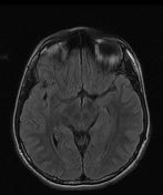
FLAIR: small area of high signal intensity at the left cerebellar hemisphere.
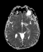
T1C+: minimal diffuse enhancement of the left optic nerve with subtle enhancement of the perineural focal lesion
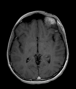
unremarkable right orbit
Case Discussion
Gliomas of the optic nerve can present as two distinct growth patterns: intraneural glial proliferation and perineural arachnoidal gliomatosis (PAG) 1. In perineural gliomatosis, there's an extraneural growth of glioma into the surrounding subarachnoid space characteristic of NF1-associated optic nerve gliomas 2. In perineural gliomatosis, you can identify the optic nerve within the lesion in contrast to the classic intraneural glioma where the tumor replaces the optic nerve 3.
The enlarged left optic nerve and perineural subarachnoid cystic dilatation, in combination with an abnormally high FLAIR signal at the left cerebellar hemisphere (FASI), highly suggest neurofibromatosis type 1 (NF1) with optic nerve glioma and perineural arachnoidal gliomatosis (PAG).


 Unable to process the form. Check for errors and try again.