
Presentation
Right conjunctival fatty lesion.
Patient Data


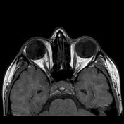
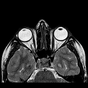
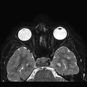
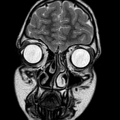
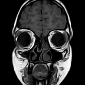
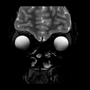
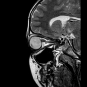
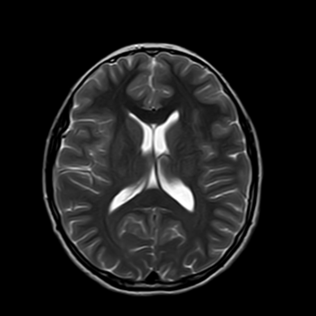
Right conjunctival epibulbar small lesion of fat signal. It measures 3 x 7 mm along its max TS and AP dimensions. It elicits a high signal on T1- and T2-weighted imaging with signal suppression on fat saturation sequences. The lesion is smoothly abutting the related lateral aspect of the globe. No connection to the intra-orbital fat.

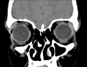
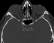
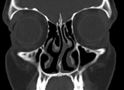
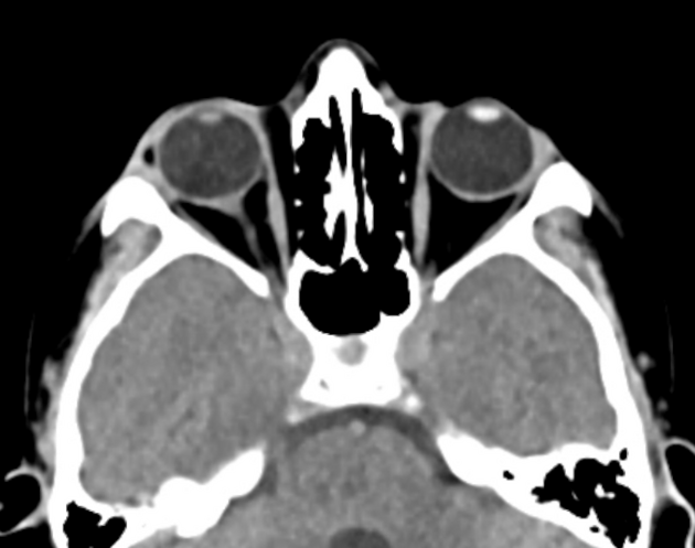
On CT, the lesion shows fat density of mean attenuation -50 HU.
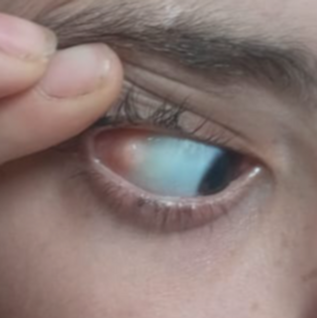
A clinical photograph of the patient shows the conjunctival fatty lesion in keeping with dermolipoma.
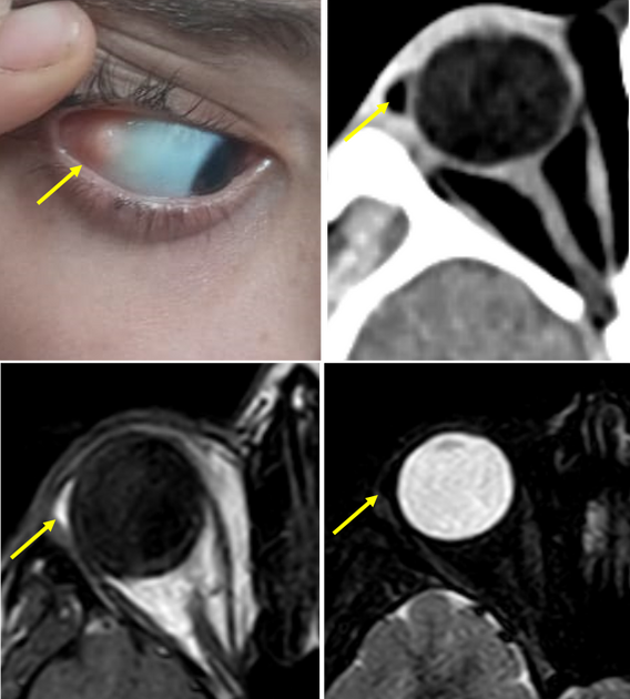
A comparison between the clinical photo and selected axial images at the same level on CT and MRI.
Case Discussion
Dermolipoma is one of the fat-containing epibulbar mass lesions of lateral canthal area beneath the temporal or superotemporal bulbar conjunctivae. This should be differentiated from subconjunctival fat prolapse as they have different radiological features and treatment strategies.
Dermolipomas are congenital and more commonly seen in young patients in contrast to the subconjunctival fat prolapse which occurs in old age. Also, dermolipoma usually has no connection to the intra-orbital fat, unlike subconjunctival fat prolapse.


 Unable to process the form. Check for errors and try again.