
Presentation
Medial foot pain. A marker was applied at the site of the patient's pain.
Patient Data
Age: 40 years
Gender: Male
From the case:
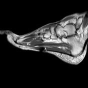
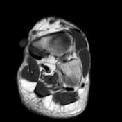
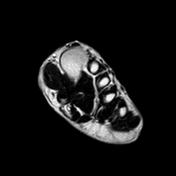
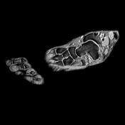
Os naviculare syndrome


Download
Info
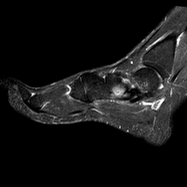
- Type II os naviculare forming pseudoarticulation with the navicular bone.
- Marrow edema signal in the opposing articular surfcaes.
- Mild tibialis posterior and flexor hallucis tenosynovitis.
- Metallic susceptibility artifacts are seen at the plantar aspect of the foot.


 Unable to process the form. Check for errors and try again.