
Presentation
Chronic focal pain over the medial aspect of the midfoot and navicular bone.
Patient Data


Findings:
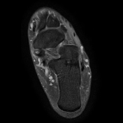
crescent-shaped accessory navicular bone (~6 x 4 x 7 mm) adjacent to the posteromedial tuberosity of the navicular bone with a fibrocartilaginous connection partly embedded within the insertion of the tibialis posterior tendon
intense bone marrow edema of the accessory navicular bone and adjacent edema of the fibrocartilaginous connection as well as mild bone marrow edema of the posteromedial aspect of the navicular tuberosity
very mild amount of fluid around the tibialis posterior tendon
Impression:
accessory navicular syndrome with stress response of the os tibiale externum (type 2)
Case Discussion
An os tibiale externum or accessory navicular bone is a relatively common accessory ossicle 1. Especially type 2, which is connected to the navicular tuberosity by a layer of fibrocartilage or hyaline cartilage, can become symptomatic and will display a typical stress response on MRI with clearly visible bone marrow edema. This is an important clue to the diagnosis of 'accessory navicular syndrome' in addition to the patient's pain being located at exactly that location.
In this case, the accessory navicular is relatively small but as opposed to the type 1, it has a semilunar shape with cartilage visible in-between itself and the navicular tuberosity.


 Unable to process the form. Check for errors and try again.