
Presentation
Advanced prostate cancer
Patient Data
Age: 80 years
Gender: Male

Download
Info

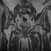
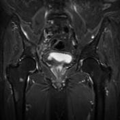
Multiple bone lesions within the pelvis, lumbar vertebrae, sacrum and femurs. These lesions are well demarcated and return high signal in STIR and low signal in T1.
From the case:
Osteoblastic metastases
Download
Info
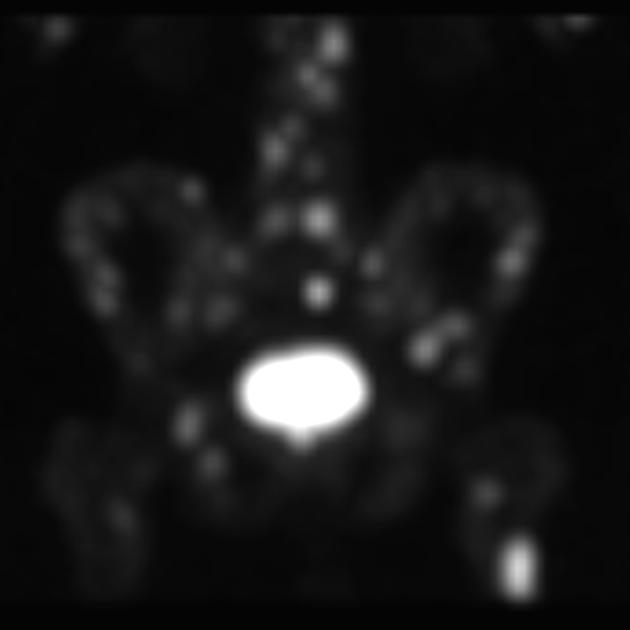
Radionuclide uptake is seen within the lumbar, sacrum, pelvic and femoral bones, corresponding to bone lesions seen in the MRI.


 Unable to process the form. Check for errors and try again.