
Presentation
Pain in the knee
Patient Data
Age: 25 years
Gender: Female
From the case:
Osteochondral defect


Download
Info
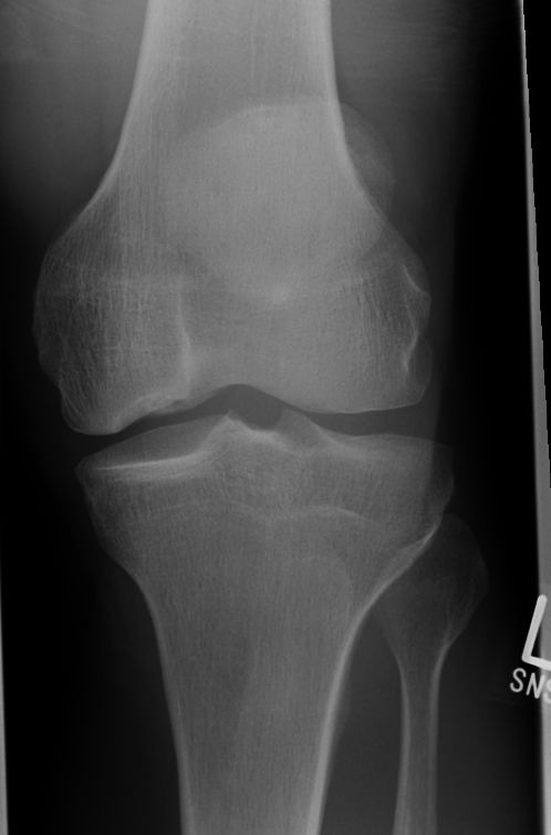
An osteochondral defect in the medial femoral condyle.
From the case:
Osteochondral defect


Download
Info

The defect was measured to be in excess of 1.2 cm and a partial tear of the ACL and edema in the medial facet of the anterior patellofemoral compartment was demonstrated.
Case Discussion
The patient presented with pain in the knee to A/E. No definite history of trauma.
Plain film showed an osteochondral defect in the medial femoral condyle.
On MRI the defect was measured to be in excess of 1.2 cm and a partial tear of the ACL and edema in the medial facet of the anterior patellofemoral compartment was demonstrated.


 Unable to process the form. Check for errors and try again.