Presentation
Pain of the right knee with intermittent locking.
Patient Data
Age: 35 years
Gender: Male
From the case:
Osteochondritis dissecans - knee





Download
Info
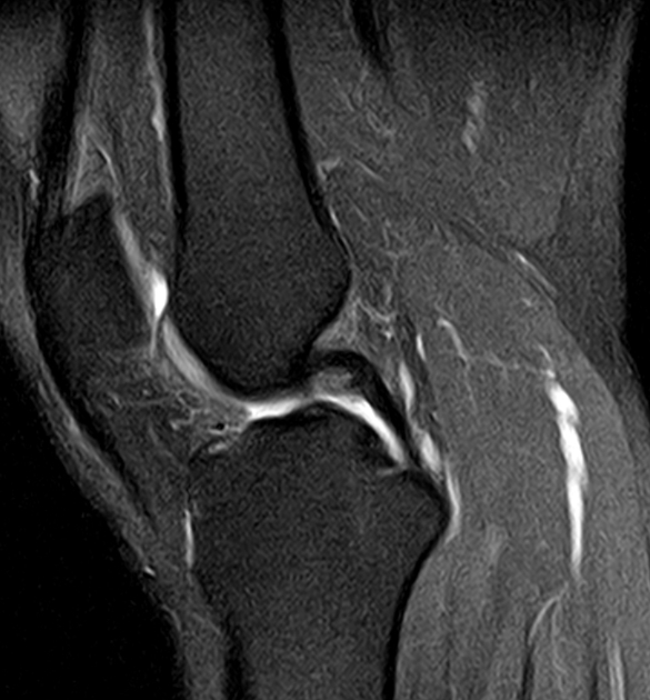
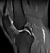
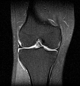
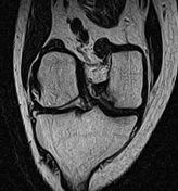
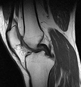
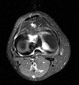
There is a well-defined osteochondral defect of the medial aspect of the medial femoral condyle with no significant underlying edema of the bone marrow. The detached osteochondral fragment is displaced, seen anteromedial to the PCL with minimal joint effusion.
ACL, PCL, and collateral ligaments are intact. No meniscal tear is seen.
Case Discussion
MRI features of an osteochondral defect with displaced detached fragment, consistent with an unstable osteochondritis dissecans of the knee, stage IV according to osteochondral injury staging.


 Unable to process the form. Check for errors and try again.