
Patient Data
Gender: Unknown
Note: This case has been tagged as "legacy" as it no longer meets image preparation and/or other case publication guidelines.
From the case:
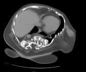
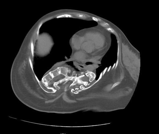
Osteogenesis imperfecta
Download
Info
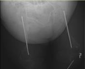
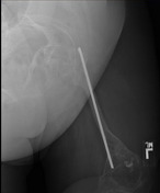
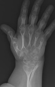
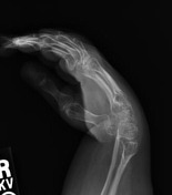
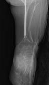
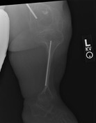
Deformed bones with severe osteoporosis showing cortical thinning.
Bilateral femoral and left tibial intramedullary nail.
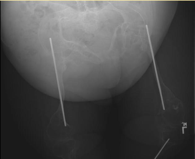
Bilateral coxa vara.
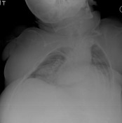
Severe thoracic kyphoscoliosis.

Case Discussion
Characteristic findings of osteogenesis imperfecta.
These images are from Dr. Paula Brill's excellent pediatric radiology collection.
Dr. Brill is a professor in the department of radiology (pediatric section) at Weill Cornell.
This case was donated to Radiopaedia.org by Radswiki.net.


 Unable to process the form. Check for errors and try again.