
Presentation
Left hip pain
Patient Data
Note: This case has been tagged as "legacy" as it no longer meets image preparation and/or other case publication guidelines.

Radiograph of the pelvis and hips of a 10 year old girl, demonstrates a lucency in the region of the greater trochanter on the left, with no convincing soft tissue mass or periosteal reaction. There is however diffuse surrounding sclerosis.


The lesion and sclerosis are confirmed on CT which demonstrates a sharply demarcated oval lesion, with a central focus of sclerosis.

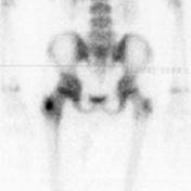
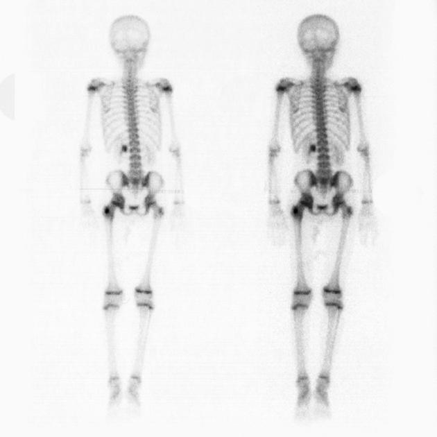
Bone scan demonstrates a double density sign, with a focal region of intense uptake, superimposed on a larger area of generalized increase activity.
Case Discussion
Findings are characteristic of an osteoid osteoma, confirmed with histology.


 Unable to process the form. Check for errors and try again.