
Presentation
Pain in her left thigh since last few months.
Patient Data
Note: This case has been tagged as "legacy" as it no longer meets image preparation and/or other case publication guidelines.
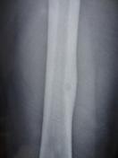
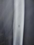
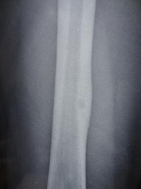
Radiograph left thigh in AP and lateral projection was done.
Radiographs show increased cortical thickness involving the mid femoral shaft on the medial aspect with associated sclerosis and a narrow zone of transition. There is alsoa focal round lucent / lytic focus within the center of the sclerotic lesion suggestive of a nidus.
Case Discussion
Features in a 12 year old patient is suggestive of an osteoid osteoma.
An osteoid osteoma is a benign bone tumor commonly seen in young individuals. It's hallmark is cortical thickening with a focal radiolucent central nidus.


 Unable to process the form. Check for errors and try again.