
Presentation
Right leg pain for a few months, exaggerated at night with no history of trauma.
Patient Data
Age: 10 years
Gender: Male
From the case:
Osteoid osteoma


Download
Info

Plain radiograph of the right leg shows focal cortical thickening and sclerosis of the lateral aspect of the proximal tibial shaft.
From the case:
Osteoid osteoma


Download
Info

Axial and coronal CT images show a small radiolucent nidus at the lateral aspect of the proximal tibial shaft with a central sclerotic dot, surrounded by reactive bone sclerosis and focal cortical thickening.
From the case:
Osteoid osteoma


Download
Info
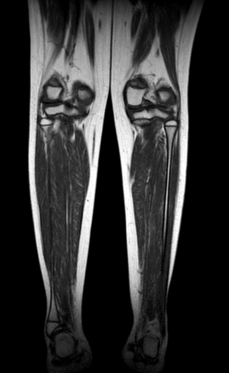
MRI demonstrates a small focus of of abnormal low signal on T1 and STIR images at the lateral aspect of the proximal tibial shaft surrounded by intense marrow edema signal.
Case Discussion
Plain X-ray, CT and MRI findings together with the clinical presentation are highly suggestive of osteoid osteoma.


 Unable to process the form. Check for errors and try again.