
Presentation
Right below knee amputation one year ago. Delayed healing of the wound at the tip of the stump.
Patient Data
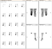

A right below knee amputation is noted. On early phase imaging, there is increased blood flow to the remaining right leg compared to the left. There is hyperemia of the stump below the knee, most marked at the distal aspect. No additional areas of hyperemia are identified on early phase imaging. Note that the whole body blood pool acquisition was terminated early before imaging the left lower leg due to patient discomfort.
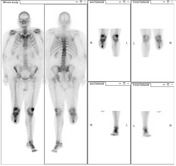
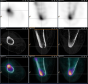
On delayed phase imaging, there is moderate to markedly increased tracer accumulation in the remnant right tibia, most focal at the distal tip. On SPECT/CT, intense tracer accumulation is present adjacent to a large cortical erosion at the cut surface of the tibia. Bone scan findings are in keeping with osteomyelitis.
There is mild to moderately increased periarticular uptake in both knees and the small joints of the left foot most in keeping with osteoarthritis.
Case Discussion
On bone scan, osteomyelitis is typically characterized by increased blood flow and hyperemia on early phase imaging, with increased tracer uptake (osteoblastic activity) on delayed phase imaging. SPECT/CT is useful to better characterized the finding, increasing the sensitivity and specificity of the diagnosis. Focal uptake in bone adjacent structural changes on CT are often encountered (e.g. periosteal reaction, cortical erosion), as in this case. CT changes in the bone may not be present, however, as bone scan abnormalities can precede CT or plain film abnormalities by some weeks.


 Unable to process the form. Check for errors and try again.