
Presentation
Fever, swelling and pain in the right thigh. Limited movement of the right hip joint
Patient Data
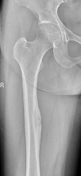

There is localized cortical thickening in the upper third of the right femur, accompanied by several areas of bone lysis. The margins of the lesions are irregular, and there is no significant periosteal reaction.
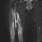

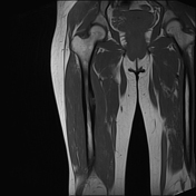
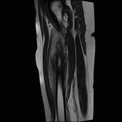
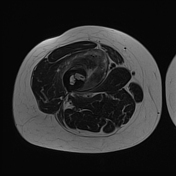
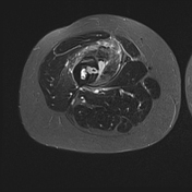
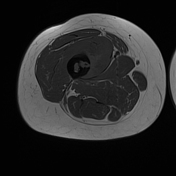

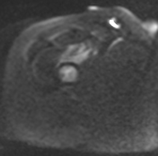
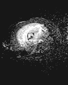

On the MRI, there is localized thickening with cortical destruction in the upper third of the right femur, suspected to contain a bony sequestrum. The lesion disrupts the cortical bone, creating a fistulous tract to the adjacent soft tissue, resulting in an abscess (diffusion restriction) in the vastus intermedius muscle.
Bone marrow edema and surrounding soft tissue swelling in the upper and middle thirds of the femur are also noted.
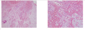
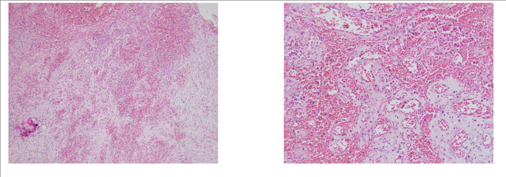
Microscopic description (from left to right)
inflamed bone tissue: normal trabecular bone structure with infiltration of inflammatory cells and hemorrhage
soft tissue around the bone: normal striated muscle structure with inflammatory fibrous tissue
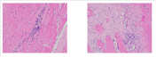
bone marrow: bone tissue with extensive infiltration of inflammatory cells
Conclusion: findings consistent with osteomyelitis
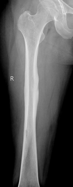
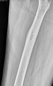
After 5 months of treatment, follow-up X-rays show a significant reduction in the lesions, with localized cortical thickening and bone sclerosis in the upper third of the femur, indicating a healing process. There are no longer any areas of bone lysis.
Case Discussion
The findings on x-ray and MRI are consistent with osteomyelitis of the femur with the formation of a sinus tract and an abscess in the adjacent vastus medialis soft tissue.
Microbiological testing of a specimen from the fluid surrounding the upper third of the femur yielded a positive result for Staphylococcus aureus. The patient was subsequently treated with abscess surgery and intravenous antibiotics. Clinical symptoms, blood tests, and imaging findings all showed significant improvement.


 Unable to process the form. Check for errors and try again.