
Presentation
Right thigh mass
Note: This case has been tagged as "legacy" as it no longer meets image preparation and/or other case publication guidelines.

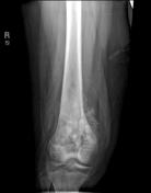

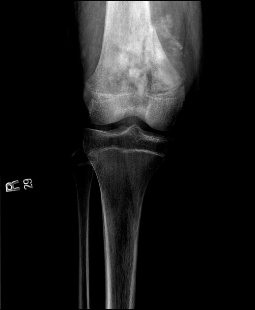
Right femur diaphyseal and metaphyseal medullary lesion with thickened cortex. It is surrounded with soft tissue swelling with irregular opacities. A linear lucent oblique line is seen traversing the femoral metaphysis medial aspect reaching articular surface, suggestive of a fracture.
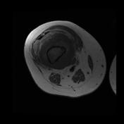
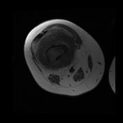
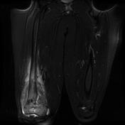
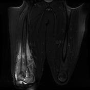
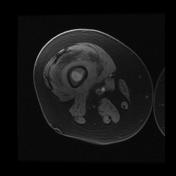
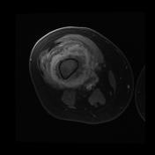
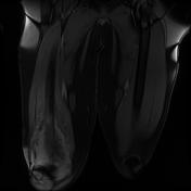
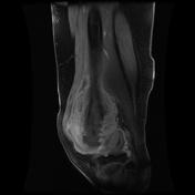
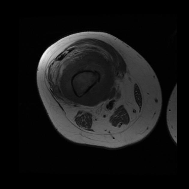
MRI of right thigh shows metaphyseal intramedullary lesion extending to lower femoral diaphysis with high signal intensity on STIR and postcontrast enhancement. It shows related cortical interruptions with surrounding soft tissue mass. Pathological fracture is seen involving the medial femoral condyle.
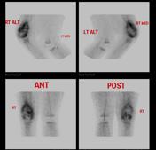
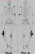
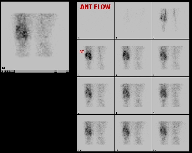
The lesion shows increased radiotracer activity on bone scan.
Case Discussion
Imaging features of osteosarcoma.


 Unable to process the form. Check for errors and try again.