
Presentation
Post-traumatic painful swelling left upper leg with difficulty in walking for one month. No discharge, fever, night sweats, anorexia or weight loss.
Patient Data

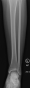
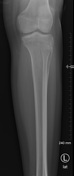
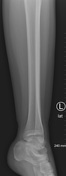
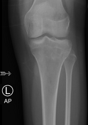
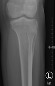
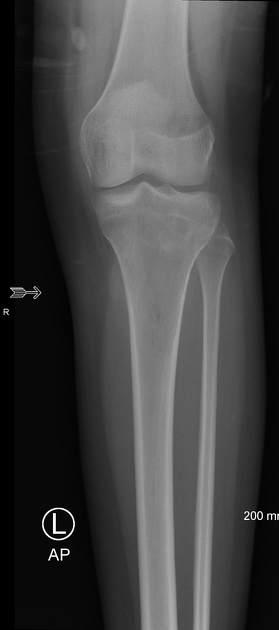
Heterogenous bone density with a subtle permeative lesion with wide zone of transition in the proximal metaphysis of the tibia. Mild cortical erosion/destruction is noted along the anterior aspect of the proximal metaphysis of the tibia. Mild soft tissue swelling along the upper anterior tibia. Morphology of the remaining tibia as well as the fibula is intact. No bone fracture or dislocation is noted.
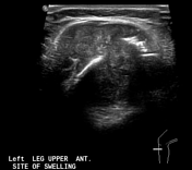
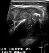
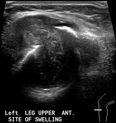
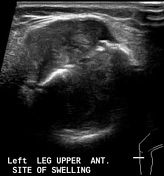
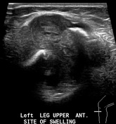
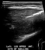
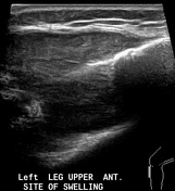
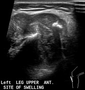
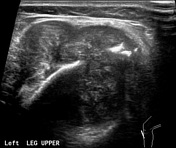
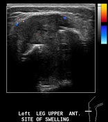
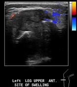
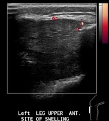

A well-defined heterogenous bilobed solid mass lesion associated with cortical destruction of the proximal anterior tibia and having intraosseous as well as extraosseous components is seen in the upper anterior left leg. No calcifications or cystic components are seen within it. Mild vascularity is seen in the periphery of this mass lesion on color Doppler ultrasound examination.


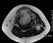
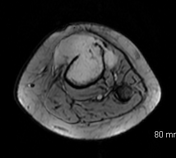
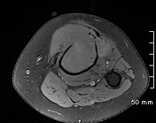
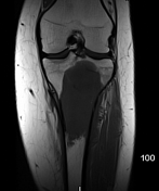
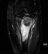
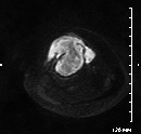
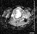
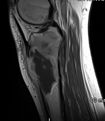
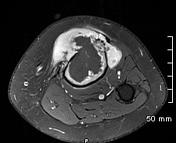
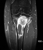
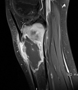
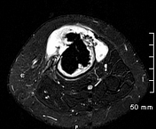

Sizable destructive lesion involving the proximal tibial metaphysis. The lesion is extending into the epiphysis through the physeal plate and associated with disruption of the anterior cortex with extension in to the surrounding soft tissues. No calcifications or hemorrhagic components are seen in it. It has solid component at the periphery and large central necrosis. The solid component shows significant enhancement as well as diffusion restriction. Fibula and knee joint are unremarkable.

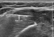
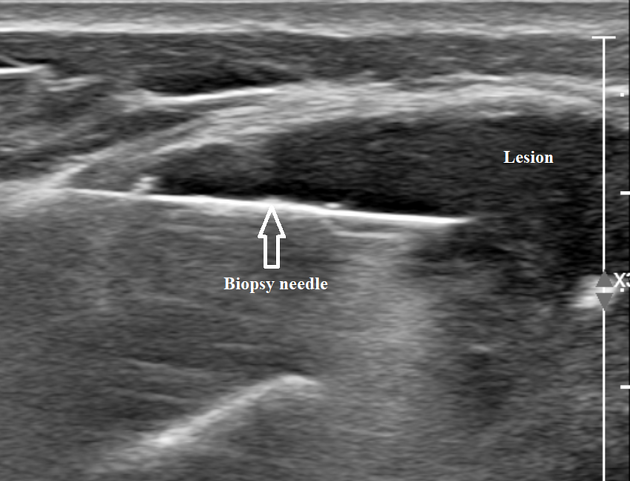
Under ultrasound guidance, using the coaxial technique, biopsy was taken from the soft tissue component of the lesion along the proximal left tibia.
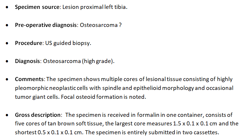
Histopathology report of the biopsy taken from the proximal left tibial lesion.
Case Discussion
Imaging features are suggestive of an aggressive/malignant bony lesion.
After consultation with an oncologic orthopedic surgeon, imaging-guided biopsy was taken and histopathology showed high-grade osteosarcoma.
Staging work up was negative for distant metastasis and the patient was referred to the medical oncologist for neo-adjuvant chemotherapy.


 Unable to process the form. Check for errors and try again.