
Presentation
Left thigh swelling.
Patient Data
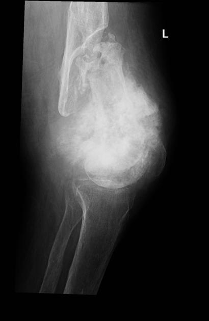
Large mass centered on the distal femur with sunburst periosteal reaction. Distal femur is sclerotic with chronic appearing distal femoral shaft pathological fracture.
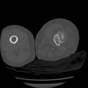

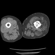

CT confirms the large distal femoral mass with large extraosseous soft tissue component and spiculated periosteal reaction. Mixed lytic/sclerotic appearance of the distal femur. Pathological fracture noted. Asymmetric muscle belly atrophy and decreased bulk in both the visualized thigh and calf.
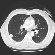
Numerous pulmonary nodules.
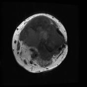
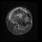
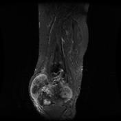

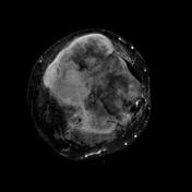
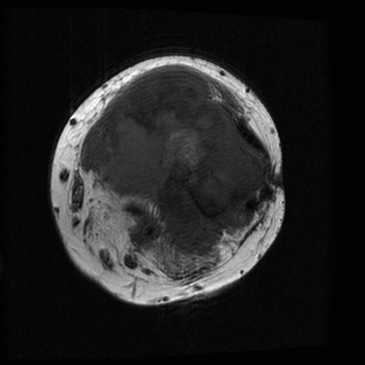
Large distal femoral mass with large extraosseous enhancing soft tissue component. Susceptibility artifact at the lateral aspect of the mass. Asymmetric muscle belly atrophy and decreased bulk in both the visualized thigh and calf.
Case Discussion
Pathologically proven osteosarcoma. Lung metastases are demonstrated.


 Unable to process the form. Check for errors and try again.